


The spine, like any structure that performs a supporting function, inevitably wears out over time. High static and dynamic loads and local overload of segments, especially the mobile upper part, lead to a decrease in regenerative abilities and gradual degeneration of cartilaginous and nearby musculo-ligament structures. By the age of 30-35, almost every person shows signs of cervical osteochondrosis to a greater or lesser extent. Although it is impossible to stop the irreversible process of biological aging, it is quite possible to slow it down.
Diagnosis
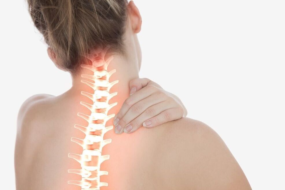
For objective assessment of the condition and detection of degenerative-dystrophic changes in the cervical spine, radiation imaging methods are used:
- plain spondylography (contrast-free X-ray study in frontal, lateral and oblique projection)
- radiograph with functional tests
- MSCT (multislice computed tomography)
- MRI
- Transparent spondylography of the upper spine is a traditional method of radiological diagnosis of cervical osteochondrosis. With its help, the condition of the vertebral bodies is assessed, their shape, height, degree of deformation and displacement towards each other are determined. X-rays visualize osteophytes, areas of enlightenment in foci of liquefaction of bone tissue.
- Spondylography with functional tests is a study aimed at identifying signs of movement disorders. X-rays are performed with a fixed maximum flexion and extension of the cervical spine.
- MSCT is a progressive alternative to X-rays. Bone structures, intervertebral discs, ligaments, spinal canal, and spinal cord are visualized in more detail in multilayer images.
- Magnetic resonance imaging allows for additional visualization of the cartilage layer and other soft tissues of the spine. The study was prescribed for severe neurological symptoms to distinguish cervical osteochondrosis from acute intervertebral hernia.
Treatment of cervical osteochondrosis
Treatment of osteochondrosis of the cervical spine is aimed at removing pain and slowing the progression of the pathological process. It is carried out in two directions: limiting the influence of adverse factors and suppressing the mechanisms of disease development.
Therapeutic and prophylactic measures that minimize the impact of the pathogen include:
- rational choice of work furniture
- use of orthopedic mattresses and pillows
- hearing, vision and posture correction
- wearing special fastening devices
- restriction of work activities associated with a long stay in a forced situation
- appropriate physical activity
- proper nutrition
There are many different methods of therapeutic correction designed to slow down the development of the degenerative process.
Massage for cervical osteochondrosis
Massage procedures aimed at relieving inflammation and relieving pain are included in the complex of mandatory therapeutic measures. The most effective types of collar massage:
- classic
- medical (manual)
- point (acupuncture)
- vacuum (canned)
- hardware
Thanks to massage techniques, the local circulation of blood and lymph is improved, tissue trophism is accelerated, muscle contractions are removed, tension in the neck is relieved, and muscle tone and elasticity are improved.
Orthopedic necklaces
Special orthopedic devices (Shants collars) are used to fix the cervical spine in the correct position. Removable structures of various sizes, shapes, and degrees of stiffness limit the usual pathological position of the head, control movement in the neck, reduce pressure on spinal segments, warm and relax tense muscles, and prevent further disease progression.
Osteochondrosis neck collar is available in several modifications:
Soft splints made of medical foamor other porous hypoallergenic materials have a notch for the chin and lower surfaces of the neck and holders. They are used to correct minor disorders in the upper spine, keep the spine in an anatomically correct position, and relax the shoulder girdle muscles.
Pneumatic collars (inflatable)they are designed to prevent pain, smooth traction, and remove vertebral artery compression.
Semi-rigid bandagesequipped with metal inserts reliably stabilize the intervertebral segments. They significantly limit the range of motion and contribute to the spread of gaps between the vertebral bodies.
Rigid corsets made of durable plasticdesigned for complete immobilization of the cervical spine in a neutral position. It is prescribed in the late stages of the disease, accompanied by compression disorders.
The necklace for osteochondrosis of the cervical spine is chosen by the doctor taking into account the age, anatomical features and stage of the degenerative process.
Manual therapy
Manual therapy aims to identify and remove blockages in motor segments. The local dosed effect on the spinal joints helps to normalize blood flow and blood supply to the brain, removing compression (pinching) and restoring the normal functioning of nerve fibers. Specific manipulations by chiropractors allow you to achieve maximum relaxation, eliminate muscle cramps, cervicogenic headaches caused by damage to the anatomical structures of the neck and tension headaches.
Acupuncture
Acupuncture, which involves the placement of acupuncture needles in bioactive sites on the neck and shoulder blades, is aimed at restoring the disturbed energy balance. By stimulating rapid contractions of sensitive nerve fibers and the release of endorphins and neurotransmitters, acupuncture for cervical osteochondrosis has a strong anti-inflammatory and analgesic effect. Thanks to this technique, numbness in the hands, dizziness, tinnitus, improves blood flow and optimizes mobility.
Physiotherapy
Physiotherapy of degenerative pathologies of the spine aims to relieve pain and stimulate the recovery process. The greatest therapeutic effect is achieved by:
- UFO
- ultrasound treatment
FAQ
How to provide help during acute pain with osteochondrosis of the lumbar spine?
In case of sudden acute pain it is necessary to repair the lower back. This will immobilize the cramping muscles and shift the load from them. Then, if possible, lay the patient on his back, placing a pillow under his bent knees. To reduce pain, you need to take a drug with analgesic and anti-inflammatory effect (NSAID). In addition, you can use an ointment or gel based on diclofenac or its analogues or put a cold compress (no more than 10 minutes). It is very important to remove the stress on the spine and visit a doctor as soon as possible.
Can lumbar osteochondrosis be done?
Physical education for lumbar osteochondrosis is not only not prohibited, but is also recommended (with the exception of periods of acute pain). However, you should be careful not to allow an axial load on your spine and categorically refuse to squat, jump and lift weights. The set of exercises should be selected by an expert on an individual basis.