


Osteochondrosis of the lumbar region is a chronic degenerative-dystrophic disease of lumbar spines that affect the structure of intervertebral discs and numerous lumbar vertebrates.It affects people of predominantly working age.It is manifested in different symptoms, of which are the main pain in the lower back and legs, limiting movements in the lower back.For diagnostics, research methods such as radiography, calculated tomography or magnetic resonance imaging of the lumbar spine are used.In this article you can more detail with the causes, symptoms and methods of diagnosing osteochondrose of the lumbar spine.
Osteochondrosis is the result of the aging of the body.These or other signs of this disease can be found in almost every person (!), Starting in 25 years.But here is the seriousness of these changes, the rate of their progress, the degree of clinical manifestations depends on many causes, primarily in the way a healthy lifestyle leads a particular person.Moderate physical activity, mandatory morning gymnastics, right pose bodies when performing multiple works (garden, construction, house and the like), orthopedic mattress are those moments that prevent the development of osteochondrose of the lumbar spine.
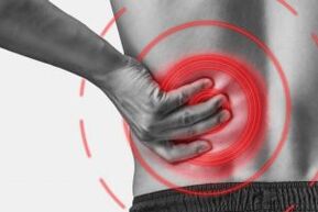
According to statistics, osteochondrosis of spine in 80% of cases is the cause of the back pain.
How is osteochondrosis develop?
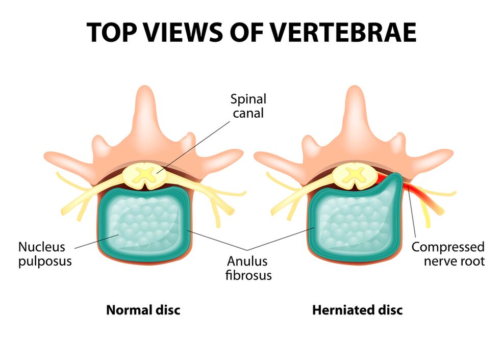
The entire spine consists of separate vertebrates, between whose bodies where there are intervertebral discs.That is, between the two vertebral is one disc.The disk consists of a gelatary (toll) core and fibrous rings.The core contains a lot of water and provides depreciation and flexibility of the spine.The own ring is located along the periphery of the core jacket, as if holding it inside.
With a long-lasting load on the core, it changes its physiological properties, lose water and dries, and at the end of the sequence: the disk is flattened, and vertebrates are approaching each other.With such processes, in the core jacket, the fibrous ring loses elasticity and, influenced by mechanical cargo, begins to protrude.This is called protrusion.Then the fibrous rings crack, and the gelatin core decreases through the resulting gaps: lively was hernia.The plot of two adjacent vertebrae and disk which is located between them, the segment of the vertebral, acquires excess mobility, which increases the load on nearby segments.The overload of neighboring segments is driven by a similar pathological process in them.These changes are called osteochondrose.
To ensure the stability of the spine, the growth of bones are formed along the edges of the vertebral bodies, increasing the area of support.This phenomenon is called spondylosis.Changes in the joints between vertebrae is called a sponding arthrosis.Usually all three pathologies - osteochondrose, spondylosis, spondila arthrosis - walk nearby.
Reasons
Why is osteochondrosis?To date there are several theories of phenomena:
- Mechanical theory: Perhaps the main reason should be considered by regular increased load on the spine.Therefore, osteochondrosis is almost mandatory fate of the driver, miners, builders and people of such professions.The occurrence of osteochondrose of the lumbar region is mainly connected to the slopes and raising weight, forced an unpleasant work pose;
- Another factor in developing is an incorrect posture, sitting in the wrong posier, which is especially important for mental workers;
- Sometimes the role plays the hereditary characteristics of the structure of the spine and the diet of his individual structures;
- Traumatic theory: Each trauma to the spine (even the most significant) can initiate a degenerative process;
- Hormonal metabolic disorders and endocrine diseases can negatively affect metabolism in the tissues of the spinal column and contribute to the development of osteochondrose;
- The age theory implies the natural spending of discs in the life process.
Rarely, only one of these theories can explain the appearance of osteochondrose in any case.More often, several factors are "blamed".
In the occurrence of osteochondrose of the lumbar spine, excess weight plays an important role, because it is itself overload for the spinal column.The higher the bodily mass index (degree of obesity), the more pronounced changes in the spine are usually.In between other reasons, causing the appearance of osteochondrose, can be noted:
- Sedentary lifestyle;
- IMORLY NUTRITION (Fast food, excess sweet, semi-refined products: All leads to imbalances of trace elements) and lack of liquid;
- Anomalies of the spine structure (for example, the presence of an additional lumbar vertebra);
- Continuous wearing shoes with a high view;
- pregnancy (due to excess load on lumbar spine);
- A sudden termination of training in people who are professionally engaged in sports;
- Smoking and alcohol abuse: As factors that speed up the aging process in the body.
Symptoms
The main manifestation of osteochondrose lumbar spine is pain.The nature of the pain, the place of phenomena and the direction of the distribution depend on which receptors are irritated, ie how much rough changes on disk and the surrounding tissues, in which the direction arises and so on.
Reflex and compression syndromes vary with osteochondrose of the lumbar spine.
Reflex syndromes are developed in cases where the receptors of the fibrous ring of the affected disk, ligaments and joints that are nearby are irritated.They are reflexive, because in addition to pain, the muscle-torture, vegetative-vascular or neuroderphic reflex changes, ie the irritation reflexes transferred to other structures, causing symptoms mainly on the side of soft tissues.
Compression syndromes occur as a result of the compression (compression) of nervous roots, blood vessels or spinal cords formed by osteochondrose changes.

Reflex syndromes of lumbar spine
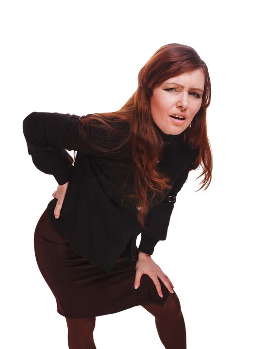
Lumbago(Feeling): Acute sudden pain in the lower back, which occurs with a clumsy movement or at the time of physical tension (much less often - without apparent reason).Lumbago is believed to be associated with the movement of the core jacket within the fibrous rings, that is, it develops in the initial stages of osteochondrose.Often pain is described as a "feeling", "the stake is stuck in the lower back."Patients are freezing in a posh in which pain caught them.The smallest move causes an increase in pain (sneezing, cough, attempt to rotate in bed, move your leg).If the person was in a sloping position at the time of Lumbago's development (which is most often happening), then it cannot be leveled.The pronounced muscle voltage in the lumbar spine appears reflexively.Along vertebrae in this area, a muscle roller feels, which is sometimes visible with naked eye without contact, and muscle tension is so spoken.I feel painful for the patient.Such an increased muscle tone performs an immobilization role, the protection of the affected lumbar segment from pathological mobility, which can cause deterioration in the state.The natural bandage spinal column in the lower back (Lordoza) is flattened, may be a curvature (scoliosis) due to muscle tension.
Lumbar- Another reflex lumbar level syndrome.This term also means the presence of pain in the lumbar area.But unlike Lumbaga, the pain does not appear acute, but gradually, within a few hours or even days.The pain is stupid, moderate intensity, is amplified during the movement, in a sitting or standing position, when moving from one position.A little relief brings the position of lying down or back with a roller under the lower back, but the passive increase in the corrected leg in this position causes increased lower back pain (Lass symptom).The palpation of the lumbar spine is painful, but the reflective tension of the muscles is less pronounced than with Lumbago, and sometimes if there is.Movements in lumbar spine are limited but possible.This means that the patient can also bend aside to a certain level (then reinforced pain).
Sciatica- Another variety of lumbar level syndrome reflex.According to this expression, the pain in the lower back, which gives the buttocks and in the leg (on the back surface).The pain is different, mostly pain, but the type of "fireplace" can occasionally appear in the leg.Just like with lumbargia, it is intensified with any movements, walking, stress, decreases lying on the back.Symptom Lasse is usually positive.The palpation of the lumbar spine is painful, as well as by pressing some points (for example, in the middle of a line that separates the buttocks from the thigh, in the middle of the back of the thigh, in the middle of the postal fossa).There is an tension of the lower back muscles.The slope forwards and on the sides are limited.
Compression syndromes of the lumbar spine
The clinical characteristic depends on which structure is subject to compression.
There are nervous roots between the vertebral hole (spinal nerves): left and right.If the pathological formations for osteochondrose of the lumbar spine (mainly discs of the discs) shine the roots, then radicalopathy is developing, whose symptoms differ for each root.Usually for all radiculopathies of the lumbar region is an increase in pain during sneezing, coughing, the lower back movements (especially tilting forward), the presence of muscular tension in the lower back.The following types of radiculopathy of the lumbar spine are most often:
- Radiculopathy L1, L2, L3: The pain occurs in the lower back, give the expected thigh.The occurrence of paresthesia is possible (a feeling of creeping goosebumps, stiffness), the surface sensitivity is disturbed (an acute touch from the usual does not differ, a feeling of cold and hot).Knee reflex declining, the weakness of the quadriceps of the thighs is revealed;
- Radiculopathy L4: The pain from the lower back gives the front of the thigh, the inside surface of the knee joint and slightly lower along the inner surface of the lower leg.In the same areas, paresthesia is felt, and the surface sensitivity is lost (reduced).Weakness is developed in the quadriceps muscles from Bedrovo, the greeting of the knee decreases;
- Radiculopathy L5: One of the frequent localizations.The pain gives buttocks, along the outer edge of the thigh, along the front surface of the lower leg to the internal edge of the foot and thumb.It feels paresthesia here, superficial sensitivity is disturbed, and here is a pain when sneezing and coughing.In addition, there is a difficulty in the extension of the thumb feet, because the muscle that performs this action was imposed by China L5.Sometimes it is difficult to stand on the fifth with exposed foot;
- Radiculopathy S1 is often located with osteochondrose of the lumbar spine.The pain gives buttocks, along the outer edge of the thigh, along the outer edge of the lower leg to the outer edge of the feet and 5. Finger, Pete.These zones are characterized by a sense of paresthesia, a reduction in sensitivity to the surface.Achilla reflex has been reduced.With damage to this spine, the weakness of the lower leg muscles and feet flops is developed, so the standing and walking of socks is difficult.
Simultaneous development of radiculopathy several roots is possible, this is especially characteristic of L5, S1.It happens that one hernia is squeezing several roots.
If the stage disc is pasted, then it can squeeze the spinal cord.It is only possible when the hernia is localized in the upper reference point, as it does not have a spinal spine (spinal roots are subjected to compression, and the spinal cord syndrome is developed, and the spinal cord syndrome is developed.
If the vessels of the lumbar regions are subjected, which conducts blood flow in the spinal cord, and then in the case of acute bloodstream disorder, a spinal stroke is developing, and with extended compression - mineopathy.Mineopathy is manifested by bilateral weakness of the leg muscles, starting with feet and gradually progress.The sensitivity in his legs is disturbed, Achilles reflex is lost, and later the knee later.It is possible to strengthen urination disorders (common, "imperative", which require immediate satisfaction, incontinence urine).
Diagnostic methods
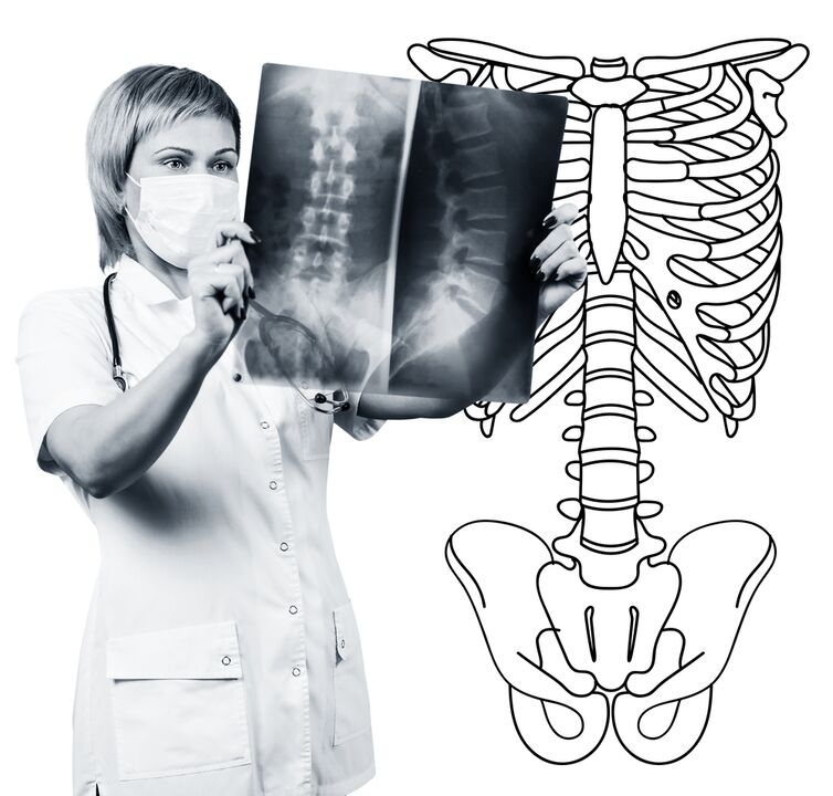
The diagnosis of osteochondrose of the lumbar spine is based on clinical data and data of additional research methods.The key role belongs to such methods as:
- Lumbar spine radiography;
- calculated tomography of lumbar spine;
- Magnetic resonance tomography of the lumbar spine.
The radiography of the lumbar spine is necessarily performed in 2 each other's vertical projections - flat rear and side.Such images allow you to see the form, contours and structure of vertebrate, height and form of intervertebral discs, abnormalities of spine and natural bandages.To display interface joints and intervertebral holes, radiograms are produced in sloping projections.In order to identify the pathological mobility of certain lumbar segments (which is a sign of osteochondrose), the radiography is performed under the conditions of the functional trial, ie in the flexion and extension of the spine.You can usually see a change in the amount of interfedgers on the front or rear in accordance with the direction of body slope, with a functional block of one of the segments, the height of the disk does not change even when bending or extension.With the pathological mobility, the displacement is determined or backward forward.The main X -Ray signs of osteochondrosis include narrowing of the intervertebral slot, pathological mobility and spine displacement, the deposition of salt in the tissue of discs (calcification), the border of the affected disk (subhondral sclerosis).The radiography of the lumbar spine is the research routine, which gradually loses significance against the background of the active implementation of new and more information research methods (CT and MRI).The radiography of the lumbar department is today used as a screening diagnostic method.
The CT lumbar spine is also done using X -Ray radiation, but the radial load on the body is much less than in X -Ray.The study is lying on the table of a special device - computer tomograph, is absolutely painless.The resulting images are processed using a computer and allow you to see significantly more structures than with spine radiography.
MRI is a method in which electromagnetic radiation is used to create images.The study is also performed in the position of lying on the table, which calls for the Chamber of Tomograph.MRI is harmless and painless.
CT or MRI lumbar spine allow you to see all spine structures, carefully examine intervertebral discs (and jacket and fibrous rings) and intervertal holes, the contents of the spinal channel.Even a slight protrusion of the interverter disc will not pass unnoticed.These methods (especially MRI) allow you to determine the direction of the disk of the disk if there are, the degree of compression of nervous roots, spinal cords.Therefore, these research methods are much informative in the diagnosis of osteochondrose of the lumbar spine from radiographs.In addition, they allow you to diagnose not only osteochondrose, but also other diseases (tumors, circulation disorders in spinal cords, abscesses, inherent in the structure of spine and spinal cord), which is important during the differential diagnosis of the causes of back pain.
Osteochondrose of the lumbar spine is a disease that most often causes pain in the back.In fact, destruction of intervertebral disks.Due to the osteochondrose of the lumbar spine, the person often loses the working capacity, because, except for pain, the disease can lead to violation of the mobility of the spine, the inability to sit, condition and walk.The symptoms of this disease are unbearable and require additional research methods to correctly confirm the diagnosis.Our father is not informative and safe modern methods of diagnosis of osteochondrose MRI spine.